
iRPE
Matériel
Cavités 10 mm, 23 mm, 36 mm, boucle de surface
Cryostat hélium liquide ESR900 Oxford instrument
Quelques points clés sur la RPE :
- Résonance Paramagnétique Electronique
- Présence d’un électron célibataire (matériaux, radicaux, métaux …)
- Technologie non invasive :
- micro-ondes 1GHz (50 mW max), champ magnétique (0,03mT) pour les échantillons de plus de 1 cm ;
- micro-ondes 10GHz (50 mW max), champ magnétique (0,3mT) pour les échantillon de moins de 1 cm ;
- temp d’expérience : 1 spectre 1-2 min,une image RPE, 10 à 100 min…
- pas de préparation spécifique de l’échantillon.
Complementary systems avaible :
- Computed X Ray microtomograph
- Gaseous anesthesia, with monitoring.
- Sample preparation Aera
- Cell culture aeera
- Quanrantine and animal facilities for mice
- X Ray CT for anatomic repositioning
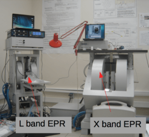 EPR platform : X-band facilities (in vitro EPR)
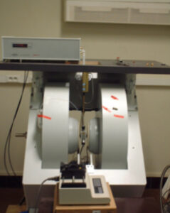
Bruker Elexsys E540 X-band EPR
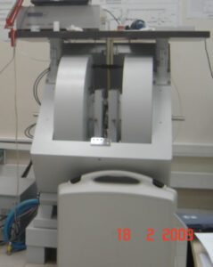
Bruker Elexsys E500 X-band EPR
Bruker ESOO and E540 X-Band EPR
- Micro-wave frequency : 1OGHz
- Magnetic Field :from 0 to 1 T
- Gradi:ent up to 0,2 mT/m
- TMS and SHQ resonator
- Stop flow for flow EPR experiments.
- Cryostat for Liquid Helium or liquid nitrogen temperature experiments.
Contacter la plateforme RPE
Sonia Lajnef, Ingénieur RPE
sonia.lajnef (a) parisdescartes.fr
01 42 86 21 75
Yves-Michel Frapart, Directeur scientifique
irpe (a) parisdescartes.fr
Accès
iRPE
CNRS UMR 8601- Université Paris Descartes
45, rue des Saints Pères
75270 PARIS Cedex 06
Spin-trapping : superoxyde and hydroxyle quantification on cells
We have a strong experience in spin trapping.
Spin trapping EPR allow quantification of superoxyde raidcal, hydroxyle radical, NO or other short lived radical using· addition of a diamagnetic species which trap the radical of interest.
Valid protocol for extracellular cell production only. Real detection on living celis.
Detection of superoxide production in stimulated and unstimulated living cells using new cyclic nitrone spin traps.
Abbas K, Hardy M, Poulhès F, Karoui H, Tarda P. Ouan 0, Peyrot F. Free Radie Biol Med. 2014;71:281-90. :

EPR spectra of DEPMPO (10 mM) + RAW mouse macrophages stimulated with PMA
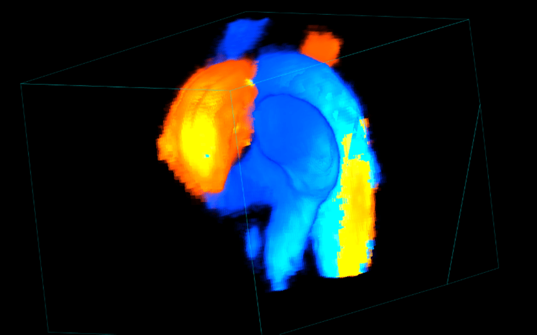
Quantification of contrast agent
Objective : Can in vivo EPR in mice help to quantify and image ultrasmall superparamagnetic iron oxide (USPIO) MRI contrast agents ?

EPR monitoring of USPIO in living mice up to 21 days after injection

Standard curve of USPIO contrast agents under in vivo EPR conditions
Conclusions
Quantification of USPIO contrast agents (P904, 046 “Dotarem”, nanotube) by in vivo EPR spectroscopy is possible. The detection limit for P904 represents 0.1µmol iron.
Good correlation between the amount of USPIO detected by EPR in ex vivo murin organs and the injected USPIO.
The width of the EPR signal is not compatible with EPR imaging.
Advanced Functional Materials, 2015, 8, p1258. DOi 10.1002/adfm.201402289, Marie H et al, Superparamagnetic Liposomes for MRI Monitoring and Extemal Magnetic Field-Induced Selective Targeting of Malignant Brain Tumors.
Nature, 2015, vol 523, p 92, doi:10.1038/nature14329, M. E. Femandez-Sanchez et al, Mechanical induction of the tumorigenic – catenin pathway by tumour growth pressure
À lire aussi
Formation pratique : échographie haute résolution du petit animal
La Plateforme Imageries du Vivant organise avec le soutien de France Life Imaging (FLI) une formation pratique en échographie pré-clinique. Cette formation est destinée aux techniciens, ingénieurs ou chercheurs souhaitant réaliser eux-mêmes des examens échographiques...
Séminaire par Craig Levin : Novel TOF PET systems under development at Standford
Ci-joint l’annonce du séminaire par Craig Levin, directeur du Molecular Imaging Program à Stanford. A 13 h, le 26 novembre au PARCC-HEGP, 56 Rue Leblanc 75015 Paris Infos pratiques : Seminaire Craig Levin
Poste d’assistant ingénieur ouvert au concours INSERM sur le Site Cochin
Un poste d'Assistant Ingénieur est ouvert au concours externe INSERM à l'Institut Cochin, sur la plateforme PIV. Vous pouvez consulter la fiche de poste et obtenir les détails à l'adresse suivante, ou consulter la fiche de poste plus bas....
Une nouvelle méthode d’imagerie développée sur la plateforme PIV passe en clinique
Le phénotype induit par les mutations du gène SDHB dans les formes graves du paragangliome (une forme rare de cancer) peut désormais être mis en évidence de façon totalement atraumatique chez les patients grâce à l'imagerie de spectroscopie par résonance magnétique....